As humans, we always strive for our major benefits. The same is true when working in a challenging scientific lab environment.
As a researcher, sitting in front of the microscope, capturing images to do a simple analysis like proliferation can prove to be tedious as well. Especially, if you need to keep a check every-time on your cell numbers at different time-intervals. In doing so, you may end-up stressing the cells. Opening the incubator several times can already make things worse for your cells.
Imagine now you can overcome these issues and avoid cell stress, thereby improving your data.
How? The zenCELLowl is here to support in your daily cell analysis!
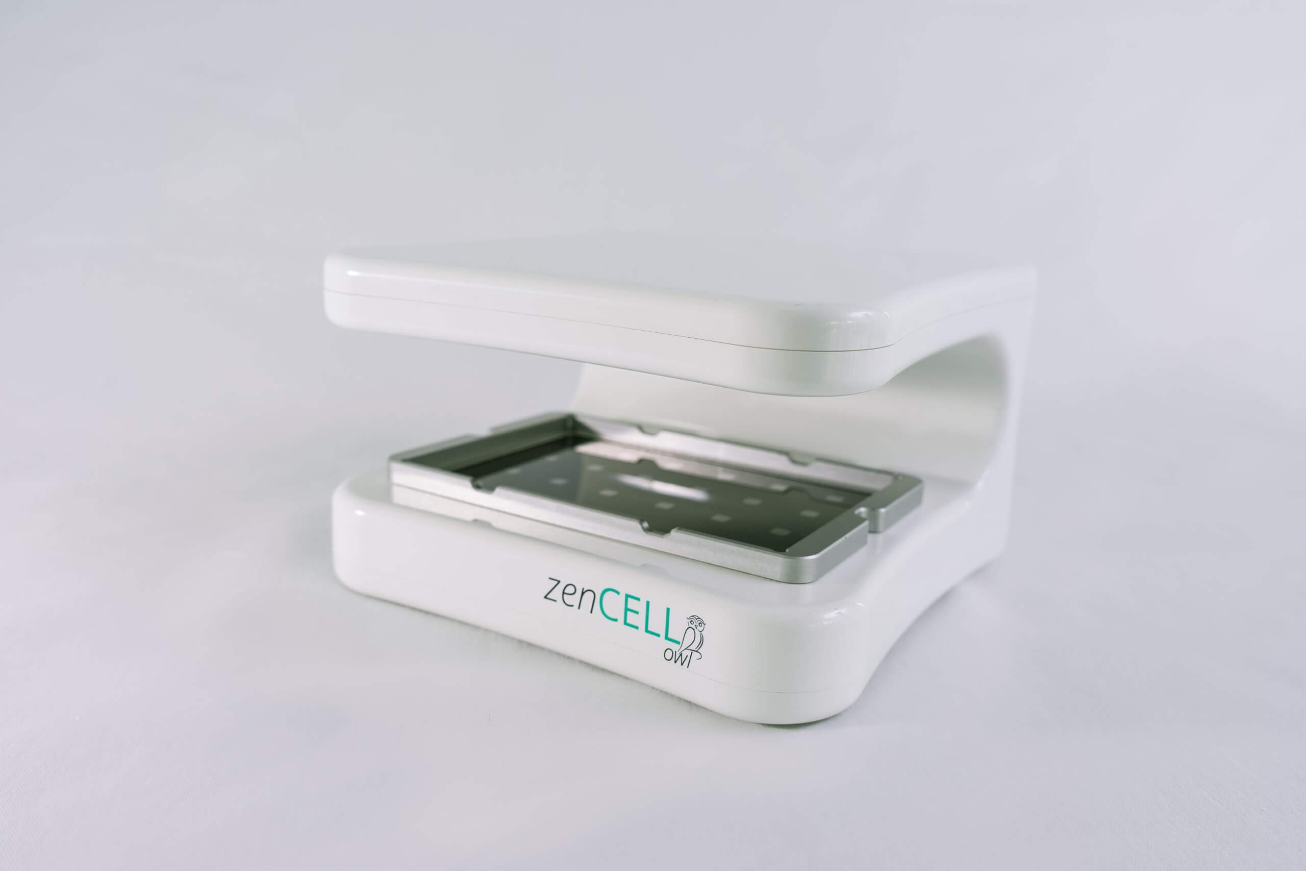
The zenCELL owl, is a 24 channels incubator microscope. It comes very handily as a compact and lightweight instrument to ensure easy use in your incubator. Cell cultures in 24 channels can easily be monitored in real-time from your PC and even remotely from home at any time (24/7).
The zenCELL owl microscope provides a comfortable and time-saving solution to monitor 3D cell culture structures such as spheroid’s size, shape, growth, adhesion, and response to external stimuli.
Due to its super small footprint, the zenCELL owl can be placed directly in the incubator, connected to the control unit, to allow automated bright-field image acquisition and real-time data without the need for user intervention.
And there is more to benefit from – the proprietary user-friendly software gives immense flexibility for operations such as time-lapse imaging, measurement of cell confluency, and number (adherent cell numbers as well as non-adherent cell number calculation).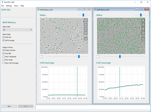
All this in 24 individual channels, making it your mini-incubator microscope.
Recently, researchers from the Institute of Analytical Chemistry, University of Regensburg, and the Fraunhofer Research Institution for Microsystems and Solid State Technologies, Munich, demonstrated a new application of time-lapse microscopy with the zenCELL owl. The researchers performed the time-lapse imaging of spheroids derived from MCF-7 cells in two different setups 1) to analyze the proliferation of spheroids in non-adhesive surface and 2) the adhesion and outgrowth of spheroids in a tissue-culture treated surface.
Overview Page for Imaging
Simple and creative solutions for every imaging need you might have. Suitable for fields such as immuno-oncology, drug discovery and diagnostics applications.
Imaging Solutions by OLS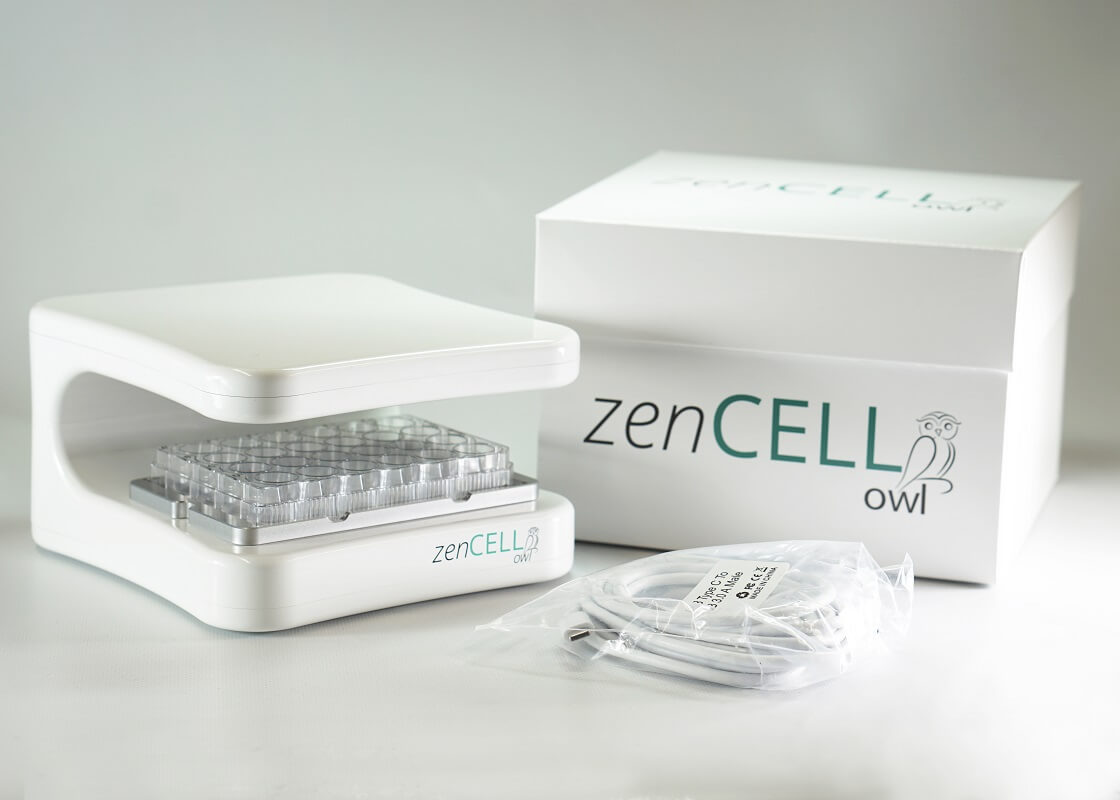
View your cell culture in real-time
Stable - Compact – Flexible Monitor your 2D and 3D cell cultures and image-based assays with an automated and simple setup.
Incubator Microscope zenCELL owl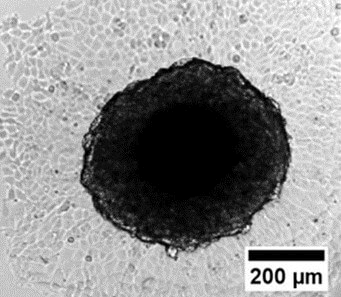
Application Note
From the research group of Prof. Dr. Joachim Wegener Institute of Analytical Chemistry, Chemo- & Biosensors, University of Regensburg
Time-Lapse Imaging of SpheroidsKarl-Ferdinand-Braun-Straße 2
28359 Bremen, Germany
OMNI Life Science GmbH
Laufenstraße 90
4053 Basel, Switzerland