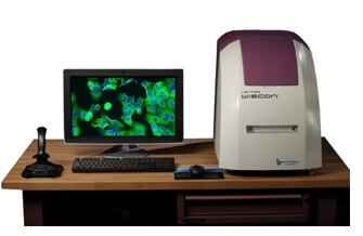
High-Content Screening Workstation WiScan® Hermes
High-quality research needs high-quality devices and equipment. Advances in cellular and molecular biology studies have revolutionized the diagnosis and treatment of many different diseases. When it comes to diagnostics, a time-efficient high-quality diagnostic solution with software for cell counting and morphology, intracellular granules, cell counts and even protein expression levels.
When it comes to drug discovery processes, they are ideally combined with modern and innovative cell imaging techniques for high-throughput screening and imaging. Advances in computational sciences and machine learning algorithms have greatly served all biological sciences and allowed greater details to be dissected at a high throughput. From microbiology to immunooncology to cell culture, OLS provides you with your ideal imaging lab partner, the Hermes WiScan® High-Content Screening Workstation.
Hermes is a cost-effective High–Content / High–Throughput Screening system that operates at extremely high speeds of image acquisition and generates very high-quality images.
Whether you are performing assay development, compound screens, transfection assays or looking at a few samples in great detail, and whether you are using 3D models (such as spheroids, organoids), zebrafish imaging, primary cells, fixed cells or live-cell imaging, WiScan® Hermes is the solution for you.
Highlights - High-Content Screening Workstation WiScan® Hermes
- Up to 7 colors + bright field
- Magnification range 2x – 60x
- Oil immersion
- Use a variety of multi-well plates, slides, dishes
- Modular platform
Take a Closer Look
Push-button operation gives access even to novice users, while experienced microscopists will appreciate the comprising application-based image analysis tools of WiSoft Athena image analysis software.
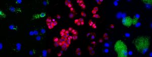
Hermes WiScan operates at extremely high speeds of image acquisition! Multi-channel imaging of a 384-well plate with four colors is finalized after 5 min – in all ranges of magnification (2x – 60x), achieving an image resolution from 0.1 – 3 µm/pixel.
Scan a 96-well plate in less than 2 min, or a 1536-well plate in 20 mins.
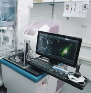
Hermes is a high-end and sophisticated modular platform, offering modular optional packages, which enable full customization for different user requirements.
Time Lapse Experiments
Live-cell imaging gets feasible by the instrument’s environmental stabilization conditions. While the objectives are moved for scanning, the microplate remains fixed during the assay.
Object Mapping
Smart object mapping further enhances throughput: at a low magnification, whole wells are scanned quickly to identify areas of interest. Then, these areas are imaged at a higher magnification (e.g., 20x – 60x) using patented automatic objective changer.
Automation
Automate your processes by letting a lab robot handle the plate stacking for most efficient, high-end, 24/7 imaging experiments, including remote accessibility. Numerous established robots are supported.
Software
Application tools for analysis and visualization comprise statistical analysis and sub-population analysis for
- Cell counting (incl. viability numbers and in colonies)
- Translocation
- Cell cycle
- Cell morphology
- Protein expression
- Intracellular granules
- Spheroids and 3D objects
- Cytometry
- Multiplexing
- Cytoskeleton fiber analysis
Flexibility
Choose from a broad range of renowned air objectives, while using a variety of plates and sample formats.
- up to 7 fluorescence colors
- photobleaching (FRAP) with high-screening throughput
- simultaneous acquisition mode for specific colors combinations
- FRET protein-interaction experiments
"The Hermes system and their new zebrafish application has been invaluable to our research, allowing us to develop a high-throughput drug screen through the fast and automated generation of data. We overcame the challenge of automating analysis of large numbers of zebrafish by collaborating with IDEA Bio-Medical to help develop an automated analysis application within their WiSoft® Athena software. IDEA Bio-Medical have been instrumental in the development of bespoke solutions that meet our needs in timely manner.”
“The Hermes system has permitted a profound increase in data acquisition for the Dent laboratory. It has permitted us to perform studies, e.g. with freshly isolated microglia, that simply could not be performed using any traditional platform. The Hermes system image analysis software has been incredibly useful in the quantification of changes in protein expression due to drug treatments. The live-cell imaging and autofocus software has enabled real-time analysis of autophagy to be performed.”
"WiScan is a great technical instrument and its image quality increases the quality of our work at the faculty of Medicine at Bratislava University. I really like WiScan. It is a great technical instrument and even though we are using it on a basic level, it gives us much better pictures compared to the fluorescent microscope which is in the next lab. And we also used it for diploma and PhD thesis, where the pictures increase the quality of the presented work.”
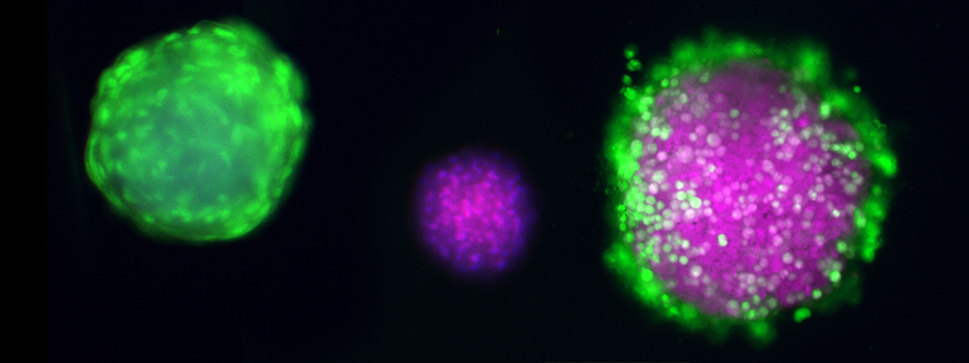
Spheroids and 3D imaging
Research in drug discovery is increasingly relying on 3D organoid and spheroid cell cultures due to their enhanced reliability as indicators of in vivo efficacy and toxicity. However, the challenges posed by the high cellular density and larger size of these cultures compared to traditional 2D cell cultures make automated image analysis during microscopy-based screening more complex.
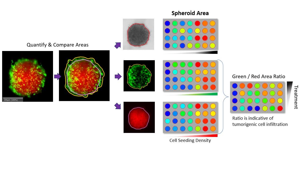
Applying robust algorithms to combined brightfield and multicolor fluorescence imaging, advanced software has been developed to morphometrically characterize each spheroid in multiple channels. This innovative approach provides insights into multiple cell types within each culture system, revealing spatial distributions of individual, categorized cells.
Utilizing widefield microscopy ensures fast acquisition and high throughput capacity, meeting the demands of large-scale drug screening trials. The resulting analyses yield multi-parametric growth curves that not only identify variations in growth patterns but also describe perturbed physiology and cytotoxicity in multiplexed assays.
Building upon these capabilities, our advanced software not only addresses the complexities associated with 3D organoid and spheroid cell cultures but also offers a comprehensive suite of features tailored to enhance every stage of the drug discovery process:
• Capture properly focused images of spheroids in their ideal growth environment in U-shape bottom plates
• Easily spot Spheroids using unique methodology of rapid scanning for spheroid localization
• Simple and labour reducing automated analysis of spheroid relevant features
• Monitor Spheroid growth over the entire plate using plate view
• Classify Spheroids of specific, desired features using sub-population tool
• Apply Live/ Dead Spheroid Assay to monitor viability of 3D tumour spheroids
• Visualize spheroid morphology over a range of depths using flexible multi-plane definitions
• Monitor Spheroid growth over the entire plate
Zebrafish Image Analysis Software
OMNI Life Science is proud to present Athena Zebrafish – the next generation in Zebrafish assays. Our new, unique dedicated analysis software for automated analysis of Zebrafish microscopy images offers simple and quick quantification of fluorescence, measurement of morphological changes & other phenotypic features in Zebrafish larvae in a high throughput format.
Athena Zebrafish is suited for a broad range of researchers and accepts multiple image format types output from nearly all microscope manufacturers.
The software permits parameter-free zebrafish analysis using simple bright-field images. It automatically detects zebrafish embryos and larvae up to 5 days old (days post fertilization), extracting the fish contour and much of its internal anatomy: yolk sac, eye, notocord, and more, along with body regions of the head, trunk, and tail.
For each of these objects, the software measures the morphology (area, length, and shape) and can detect fluorescence in associated color channels. Both fluorescence intensity and spot/structure detection within specific anatomy are supported.
Download a free trial of the Zebrafish Software
Rare events
Upcoming WiScan Hermes events
Currently there are no upcoming events. Have a look at our overview of all events.
Downloads
WiScan Brochure
WiScan Brochure
Athena Software Flyer
Athena Software Flyer
Hermes 24/7 Flyer
Hermes 24/7 Flyer
Hermes Zebrafish Screening Flyer
Hermes Zebrafish Screening Flyer
Hermes Product Family Brochure
Hermes Product Family Brochure
Hermes With Oil Objectives Brochure
Hermes With Oil Objectives Brochure
Contact
Karl-Ferdinand-Braun-Straße 2
28359 Bremen, Germany
OMNI Life Science GmbH
Laufenstraße 90
4053 Basel, Switzerland